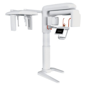
Dental CBCT provides three-dimensional imaging of teeth, jaws and other oral and maxillofacial structures.
Unlike conventional 2D dental radiography, cone beam computed tomography captures volumetric information that can be reconstructed into 3D images and examined from different planes.
This can be useful when a clinical question requires anatomical information that may not be adequately answered by a conventional 2D image.
A CBCT machine is therefore not simply a higher-resolution replacement for every dental X-ray.
The appropriate imaging method depends on the diagnostic question, patient factors and clinical requirements.
Current dental imaging guidance emphasises that CBCT should be used when clinically justified and when lower-exposure imaging is unlikely to provide the information required.
For dental clinics, implantologists, oral surgeons, orthodontists, hospitals and imaging centres, purchasing a CBCT system requires consideration of image quality, field of view, voxel size, workflow, software, physical space, operating requirements and ongoing support.
AGKEM’s CBCT category currently features the Fussen Dentrix 50 CBCT for professionals researching 3D dental imaging equipment.
Dental CBCT, or cone beam computed tomography, is a 3D X-ray imaging technology used to capture volumetric information about dental and maxillofacial structures.
During a CBCT scan, the imaging system rotates around the patient and captures multiple X-ray projections. Software then reconstructs these data into a three-dimensional image.
The resulting volume can be reviewed in different planes rather than as a single 2D projection.
This can help dental professionals examine teeth, roots, jaws and other relevant anatomical structures in situations where three-dimensional information is clinically useful.
CBCT images are commonly represented using voxels, which are three-dimensional elements of image data.
The voxel size is one of the specifications buyers may encounter when comparing CBCT systems, but it should not be considered in isolation.
The appropriate combination of voxel size, field of view, image quality and exposure settings depends on the intended clinical application.
A CBCT scan can be useful for a range of dental and maxillofacial applications when three-dimensional information is clinically indicated.
CBCT may support implant-related planning by providing 3D information about the relevant jaw anatomy.
This can be useful when the clinician needs spatial information that a conventional 2D image cannot adequately provide.
Three-dimensional imaging can be considered for selected endodontic cases where additional anatomical information may assist assessment or treatment planning.
CBCT may help clinicians assess the relationship between teeth and surrounding anatomical structures when planning certain surgical procedures.
Three-dimensional imaging can be useful when evaluating the position and anatomical relationship of impacted teeth, particularly where 2D images do not provide sufficient information.
In selected orthodontic cases, CBCT can provide 3D information about craniofacial and dental structures.
It should be used when the additional information is clinically justified rather than as a routine replacement for 2D imaging.
CBCT may provide useful 3D anatomical information for selected examinations involving the jaws and surrounding structures.
Depending on the clinical question and imaging requirements, CBCT may be considered when bony TMJ structures need three-dimensional evaluation.
CBCT data may be used for selected airway-related assessments. The exact role of CBCT depends on the clinical indication and the professional’s diagnostic approach.
For complex cases, 3D imaging can provide additional anatomical context that may be relevant to treatment planning.
The important principle is that CBCT should be selected because it answers a specific clinical question, not simply because it provides more information.
The ADA and FDA recommend considering whether alternative imaging can provide the required information with less radiation exposure.
CBCT and OPG, or panoramic X-ray, serve different imaging purposes. CBCT provides 3D volumetric information, while OPG produces a 2D panoramic representation of the jaws and teeth.
Feature | CBCT | OPG / Panoramic X-Ray |
Imaging | 3D volumetric imaging | 2D panoramic imaging |
Anatomical view | Multiple planes and 3D reconstruction | Broad 2D view of the jaws and teeth |
Image data | Volumetric data using voxels | 2D image data using pixels |
Typical use | Selected cases requiring 3D information | General panoramic assessment and other appropriate indications |
Implant assessment | Can provide 3D anatomical information when indicated | Provides 2D information |
Impacted teeth | Can show 3D relationships | Provides a panoramic overview |
Radiation considerations | Exposure varies by protocol and system | Exposure varies by equipment and protocol |
Clinical choice | Used when 3D information is clinically justified | Often suitable when a 2D image answers the clinical question |
CBCT is therefore not universally better than OPG. The correct modality depends on what the dentist or specialist needs to evaluate.
Current ADA recommendations specifically emphasise selecting 2D or 3D imaging according to the individual clinical scenario.
The main value of 3D dental imaging is the additional spatial information it can provide in appropriate cases.
A CBCT scan provides a volumetric representation rather than a single 2D projection, allowing clinicians to examine structures from different planes.
CBCT datasets can be reconstructed and reviewed in multiple planes, which may help when anatomical relationships need to be assessed from more than one direction.
Three-dimensional information can contribute to planning for selected implant, surgical, endodontic, orthodontic and other complex cases.
CBCT can show relationships between structures that may be difficult to appreciate on a conventional 2D image.
CBCT produces digital imaging data that can be stored electronically as part of an appropriate clinical records system.
3D images can provide a visual aid when discussing relevant anatomical findings and treatment considerations with patients or other dental professionals.
CBCT can form part of a broader digital workflow involving treatment planning, digital records and, where compatible systems are used, other forms of digital dentistry.
These benefits do not mean that every patient requires CBCT. The additional information should have a clear clinical purpose.
Yes, CBCT uses X-rays and therefore involves exposure to ionising radiation. Radiation exposure varies according to the machine, scan protocol, field of view, exposure settings, patient size and the anatomical region being examined.
Dental professionals should use CBCT when clinically justified and select protocols that provide adequate image quality for the diagnostic task while keeping exposure as low as reasonably achievable.
The FDA recommends justification and optimisation principles for dental CBCT, including selecting appropriate technique factors for the clinical indication and patient.
The ADA’s updated 2026 guidance similarly recommends using CBCT only when clinically necessary and considering lower-exposure alternatives where they can provide the required information.
Choosing a dental CBCT machine requires evaluating the complete system rather than one headline specification.
Review the manufacturer’s published image-quality specifications and consider whether they fit the examinations your clinic expects to perform.
Field of view determines the volume that can be captured during an examination. A system offering more than one FOV may provide flexibility for different clinical requirements.
Voxel size describes the size of the three-dimensional image elements. Smaller voxel options can be relevant for examinations requiring finer spatial detail, but the appropriate setting depends on the clinical indication and exposure considerations.
Check both exposure time and image reconstruction time. Workflow efficiency depends on the complete process, not only the duration of X-ray exposure.
Review the available image-reconstruction and review functions. Software should be practical for the team using the machine and appropriate for the intended clinical workflow.
Consider how the machine accommodates different patients and whether its positioning process is clear and repeatable.
CBCT systems require dedicated physical space. Measure the room and account for the machine dimensions and recommended installation space before purchasing.
Confirm what image data formats are supported and whether DICOM export is available where it is required for your clinic, referral or digital workflow.
If the clinic already uses intraoral scanning, digital impressions, treatment-planning software or CAD/CAM systems, assess how CBCT data will fit into the wider workflow.
Compatibility should always be confirmed rather than assumed.
Ask about installation, training, warranty, technical support, preventive maintenance and spare-parts availability.
These factors can affect the practical value of a CBCT machine in India over its ownership period.
The CBCT machine price can vary significantly because systems differ in imaging specifications, field of view, detector technology, software, accessories and configuration.
The total investment may also be affected by:
AGKEM’s current Fussen Dentrix 50 product page uses an “Enquire Now” process and does not display a fixed public selling price.
Dental professionals should therefore contact AGKEM for a quotation based on the required configuration rather than relying on an unverified online price.
A dental CBCT machine may be worth evaluating for practices that regularly handle cases where 3D information provides meaningful clinical value.
Clinics with significant implant-related work may consider CBCT as part of their diagnostic and treatment-planning infrastructure where clinically indicated.
Practices handling complex surgical and maxillofacial cases may have greater need for 3D anatomical imaging.
A multi-specialty clinic may benefit from having access to 3D imaging for selected cases across different departments.
Hospitals and dental colleges may consider CBCT based on their patient mix, teaching requirements, specialist services, equipment infrastructure and expected utilisation.
Imaging centres serving dental professionals may evaluate CBCT according to referral volume, service requirements and the range of examinations they intend to support.
Smaller clinics should consider the same factors carefully. Patient volume, clinical requirements, available space, capital investment, staffing, service arrangements and expected utilisation all matter when determining whether owning a CBCT machine is practical.
CBCT can become one component of a connected digital dentistry workflow.
A typical process may involve:
Patient Assessment → Clinically Indicated Imaging → CBCT Acquisition → 3D Reconstruction → Image Review → Treatment Planning → Digital Record / Referral
Depending on the systems used by a clinic, CBCT data may also form part of workflows involving implant planning, digital impressions, intraoral scanning, CAD/CAM processes or dental laboratory communication.
However, software compatibility should always be verified for the exact systems being used.
A CBCT machine should not be assumed to integrate directly with every intraoral scanner, CAD/CAM platform or planning application.
For clinics building a broader digital ecosystem, AGKEM also lists Intraoral Scanner and Dental X-ray / RVG equipment within its dental clinic equipment categories.
AGKEM’s current CBCT category lists the Fussen Dentrix 50 CBCT as its available CBCT product.
The Fussen Dentrix 50 is described on AGKEM’s official product page as a 2-in-1 or 3-in-1 CBCT imaging system, with CBCT and panoramic imaging and optional cephalometric capability depending on configuration.
The page also lists AI-based image review functions and cloud functions for reviewing and sharing 3D images.
AGKEM lists the following specifications for the Fussen Dentrix 50:
Specification | Published Information |
Tube voltage | 60 to 90 kV |
Tube current | 4 to 10 mA |
FOV | 15 × 9 and 5 × 5 |
Focal spot | 0.5 mm |
Grey scale | 16 bit |
Voxel options | 0.25, 0.2, 0.1 and 0.075 mm |
Total filtration | 2.5 mm Al |
Scan position | Standing and wheelchair |
Exposure time | ≤15 seconds |
Reconstruction time | <45 seconds |
Configuration | 2-in-1 and 3-in-1 options |
Recommended installation space | Varies by configuration |
These specifications are published by AGKEM and should be evaluated in relation to the clinical examinations, room configuration and workflow of the purchasing organisation.
The product page also describes intelligent image-processing functions intended to address image quality, noise and artefacts, along with user-oriented patient positioning.
These are manufacturer-described features and should be evaluated alongside practical workflow requirements before purchase.
CBCT in dentistry is a 3D X-ray imaging method that creates a volumetric representation of dental and maxillofacial structures.
The scanner rotates around the patient to collect X-ray data, which is reconstructed by software into a 3D image that can be examined in different planes.
CBCT stands for Cone Beam Computed Tomography. It is a form of X-ray imaging in which a cone-shaped X-ray beam is used to acquire data that can be reconstructed into three-dimensional images of dental and maxillofacial structures.
A dental CBCT scan is used when three-dimensional imaging can provide clinically relevant information.
Applications can include selected implant planning, endodontic assessment, oral surgery, impacted teeth, orthodontic assessment and evaluation of jaw structures.
The examination should be based on a specific clinical need rather than performed routinely.
CBCT provides 3D volumetric information, while an OPG provides a 2D panoramic image.
CBCT can show anatomical relationships from multiple planes, whereas OPG is useful when a broad 2D overview answers the clinical question.
Neither modality is universally preferable, so selection should depend on the diagnostic requirement.
CBCT is not automatically better than an OPG because the two modalities provide different types of information.
CBCT can be useful when 3D anatomy is needed, while OPG may be sufficient for many situations that require a panoramic 2D view.
Current guidance supports choosing the modality according to clinical need.
Yes, dental CBCT uses ionising radiation because it uses X-rays. Exposure varies by machine, field of view, scan protocol, patient size and other technical factors.
CBCT should be used when clinically justified, with settings optimised for the diagnostic task.
CBCT scan time depends on the machine and selected protocol. The exposure itself may be relatively brief, but the complete workflow also includes patient positioning and image reconstruction.
For the Fussen Dentrix 50, AGKEM publishes an exposure time of up to 15 seconds and reconstruction time of less than 45 seconds.
CBCT can provide detailed three-dimensional anatomical information, but image quality depends on the system, protocol, voxel size, positioning and other factors.
Accuracy should not be reduced to a single specification. Clinicians should evaluate whether the selected system provides appropriate information for the examinations they intend to perform.
CBCT can be useful for selected implant-planning cases because it provides three-dimensional information about relevant jaw anatomy.
It may help clinicians assess spatial relationships that are difficult to determine from 2D imaging.
The decision to use CBCT should still be based on the individual clinical requirement.
CBCT may be used in selected endodontic cases when additional 3D information is clinically useful.
It can provide views of dental structures that may not be adequately represented on conventional 2D radiographs.
It should not be treated as a routine requirement for every root canal case.
Yes, CBCT can show the three-dimensional position of impacted teeth and their relationship with surrounding structures.
This can be useful when conventional radiographs do not provide enough spatial information for the clinical question.
The need for CBCT should be determined by the treating dental professional.
CBCT is a cone-beam form of computed tomography designed for applications involving regions such as the teeth and maxillofacial structures.
Conventional medical CT uses a different acquisition geometry and is generally designed for broader medical imaging applications.
They should not be considered interchangeable simply because both produce 3D images.
Before buying a CBCT machine, evaluate image quality, field of view, voxel options, scan and reconstruction time, software, patient positioning, machine footprint, data export, DICOM support, workflow integration, installation, warranty, service and total cost of ownership.
The specifications should match the clinical applications and expected utilisation of the facility.
CBCT machine price in India varies according to the system's imaging capabilities, configuration, software, accessories and support arrangements.
Installation, training, warranty and service can also affect the total investment. AGKEM's Fussen Dentrix 50 page currently uses an enquiry-based purchase process rather than listing a fixed public price.
Field of view, or FOV, is the volume of anatomy captured during a CBCT examination. A smaller FOV can focus on a limited area, while a larger FOV can encompass more anatomy.
The appropriate FOV depends on the clinical question and should be selected according to the required diagnostic information.
Voxel size refers to the dimensions of the three-dimensional elements that make up a CBCT volume.
Smaller voxel options can provide finer spatial sampling, but the appropriate choice depends on the clinical task, image quality requirements and exposure considerations.
AGKEM lists multiple voxel options for the Fussen Dentrix 50.
CBCT data can form part of a digital dentistry workflow where the imaging system and other software support the required data exchange.
Potential applications include implant planning, digital treatment planning and selected CAD/CAM-related workflows.
Compatibility should always be verified for the specific CBCT and software systems being used.
Dental clinics that regularly handle cases requiring 3D imaging may have stronger reasons to consider a CBCT machine.
This can include implant-focused practices, oral surgery practices, multi-specialty clinics, hospitals and imaging centres.
Smaller clinics should evaluate patient volume, clinical requirements, space, investment and service support before purchasing.
CBCT is often part of a wider dental imaging equipment ecosystem. Depending on clinical requirements, dental professionals may also evaluate complementary technologies available through AGKEM.
AGKEM’s current Dental Clinic equipment navigation includes CBCT, Dental Chair, Dental X-ray / RVG and Intraoral Scanner categories, allowing practices to evaluate imaging and other clinical equipment as part of a broader setup.
For a clinic evaluating a dental CBCT machine, the most important question is not simply which system has the highest specification.
The better approach is to match the machine’s imaging capabilities, workflow, physical requirements and support arrangements to the clinical questions your practice actually needs to answer.
For current specifications and a quotation for the Fussen Dentrix 50, review the official AGKEM product page and contact AGKEM with your requirements.