Dental X-ray equipment plays an important role in diagnosis and treatment planning when radiographic imaging is clinically indicated. From routine intraoral radiographs to digital RVG systems, the right imaging setup can help a dental practice capture, view and manage radiographic information as part of its clinical workflow.
Modern dental practices can choose between different types of X-ray equipment and image receptors depending on the examinations they perform. A basic intraoral X-ray setup may include an X-ray generator and digital intraoral sensor, while larger practices may also use panoramic or CBCT systems for broader imaging requirements.
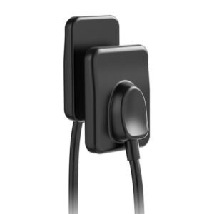
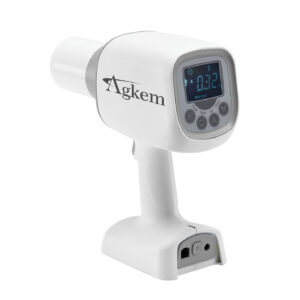
AGKEM offers dental imaging equipment including dental X-ray solutions and the PhoCo Intraoral Sensor as part of its clinical equipment range. Its Dental X-Ray/RVG category currently includes the PhoCo Intraoral Sensor and Portable Dental X-Ray Machine Gun-Type.
A dental X-ray machine produces a controlled beam of X-rays that passes through oral and maxillofacial structures to create a radiographic image.
Different examinations require different imaging techniques. An intraoral X-ray focuses on a relatively small area and places the image receptor inside the patient’s mouth. Other systems, such as panoramic X-ray and CBCT, are designed for wider or three-dimensional imaging.
The choice of equipment should therefore be based on the diagnostic information required rather than simply choosing the highest-specification machine available.
Radiographs should be prescribed based on the patient’s individual clinical needs and the information required for diagnosis or treatment planning.
RVG is commonly associated with RadioVisioGraphy and is widely used in dentistry to describe digital intraoral radiography.
An RVG sensor replaces conventional dental X-ray film as the image receptor. The sensor is positioned inside the patient’s mouth during exposure and captures the X-ray information digitally. The resulting image can then be displayed on a connected computer or imaging system.
Unlike conventional film, digital imaging does not require chemical processing before the image can be viewed.
The digital workflow can also make it easier to store radiographs electronically and incorporate them into appropriate patient-record systems.
A typical digital intraoral radiography workflow involves several components:
X-Ray Generator → Intraoral Sensor → Image Acquisition → Imaging Software → Digital Image
The X-ray generator produces the radiation required for the examination. The RVG sensor captures the X-ray information after it passes through the relevant anatomical structures. The imaging system then processes the captured information into a digital radiograph.
The quality of the final image depends on more than the sensor alone. Positioning, exposure settings, the X-ray generator, sensor characteristics, software and operator technique all contribute to image quality.
Dental X-ray equipment can be broadly divided according to the type of examination and image produced.
Intraoral X-ray systems are used for examinations where the image receptor is placed inside the patient’s mouth.
They can be used for different types of intraoral radiographs, including periapical, bitewing and occlusal imaging, depending on the clinical requirement.
A digital RVG sensor can be used instead of conventional film to capture these images.
Portable dental X-ray units are designed to provide greater flexibility in situations where a fixed installation may not be practical.
AGKEM currently lists a Portable Dental X-Ray Machine Gun-Type in its Dental X-Ray/RVG category.
When considering a portable X-ray system, practices should evaluate its intended use, regulatory requirements, radiation-safety provisions, operating procedure and compatibility with the existing imaging workflow.
Panoramic imaging captures a broader view of the jaws and dentition than a typical intraoral radiograph.
It can be useful when a larger anatomical overview is required. However, a panoramic image and an intraoral radiograph answer different imaging needs and should not be treated as interchangeable.
CBCT creates three-dimensional imaging data and can provide information that cannot be obtained from a conventional two-dimensional dental X-ray.
CBCT may be considered when three-dimensional information is clinically necessary, such as in selected implant planning, complex endodontic assessment, oral surgery and other applications.
AGKEM maintains a separate CBCT category within its dental clinical equipment range.
Dental X-ray and RVG are related but do not mean exactly the same thing.
A dental X-ray refers to the radiographic imaging process and equipment used to generate X-ray images.
RVG generally refers to the digital image-receptor system used to capture an intraoral radiograph.
For example, a dental clinic may have an intraoral X-ray generator connected to a computer-based workflow using an RVG sensor.
In simple terms:
Component | Main role |
Dental X-ray generator | Produces the X-ray beam |
RVG sensor | Captures the X-ray information digitally |
Imaging software | Processes and displays the image |
Computer/display | Allows the image to be reviewed and stored |
All of these components contribute to the overall digital radiography workflow.
A digital intraoral sensor can be used when an intraoral radiograph is clinically indicated.
Depending on the examination, intraoral radiographs may contribute to:
The sensor itself does not diagnose a condition. It captures the radiographic image, which must then be interpreted by a qualified dental professional alongside the clinical examination and other relevant information.
One of the practical differences between digital sensors and conventional film is the ability to view the image electronically without the chemical film-processing stage.
This can simplify the imaging workflow and allow the dental team to review the radiograph during the appointment.
Digital radiographs can be incorporated into electronic records and stored within an appropriate imaging or practice-management workflow.
This can reduce reliance on physical film storage and make previous images easier to retrieve when required.
Digital imaging software may provide functions such as magnification, brightness and contrast adjustment.
These tools can help clinicians review an image from different display perspectives. They do not, however, replace the need for correct positioning and appropriate image acquisition.
Digital images can be shared through appropriate clinical systems when consultation or referral is required.
This can be useful when a dentist needs to communicate radiographic findings with another dental professional.
Displaying a radiograph on a monitor can make it easier to explain relevant findings to patients and discuss treatment considerations.
Digital imaging can offer opportunities to reduce radiation exposure compared with some conventional film workflows, but it would be inaccurate to assume that every RVG system automatically results in a particular radiation dose.
Actual exposure depends on factors such as:
Radiographs should be clinically justified and obtained using appropriate exposure protocols.
The objective should be to obtain the diagnostic information required using appropriate imaging technique and radiation-safety practices.
An RVG sensor is a clinical device that may be used repeatedly throughout the working day, so specifications such as resolution are only one part of the buying decision.
Sensor dimensions can affect positioning and patient comfort.
A sensor intended for routine adult imaging may have different dimensions from a smaller sensor designed for patients with smaller mouths.
AGKEM’s PhoCo range includes different sensor sizes, including PhoCo Mini and PhoCo Pro. The product information lists the Mini at 20 mm × 30 mm and the Pro at 25 mm × 30 mm.
Resolution is an important specification when comparing digital sensors. AGKEM lists 12 to 14 lp/mm for its PhoCo sensor.
However, resolution should not be considered independently from exposure technique, positioning, software and the overall imaging system.
Check how the sensor connects to the computer and whether its software works with the clinic’s existing setup.
AGKEM’s published PhoCo specifications include USB 2.0 connectivity and support for BMP, JPEG, PNG, TIFF and DICOM image formats.
Because an intraoral sensor is used in a clinical environment, protection against moisture and appropriate cleaning and disinfection procedures are important considerations.
AGKEM lists an IP68 rating for the PhoCo sensor.
Always follow the manufacturer’s instructions for barrier protection, cleaning, disinfection and handling.
The sensor cable is another practical consideration. Repeated bending, pulling or improper handling can affect equipment life.
When comparing RVG systems, consider how the sensor and cable will be handled during routine clinical use.
An RVG sensor should fit into the clinic’s existing computer and imaging workflow.
Before purchasing, check operating-system requirements, image formats, software compatibility and whether the system can be integrated with the practice’s existing records or imaging setup.
The right dental X-ray machine depends on the type of imaging the clinic performs and how the equipment will be used.
Start with the types of radiographs routinely required in the practice.
A clinic primarily performing routine intraoral imaging may have different requirements from a multi-speciality practice that also needs panoramic or three-dimensional imaging.
Review the manufacturer’s specifications for the X-ray generator and ensure the equipment is appropriate for the intended examinations.
If you are building a digital intraoral workflow, determine whether you will use an RVG sensor or another digital receptor.
Fixed and portable systems have different practical requirements. Consider the available space, installation requirements and how the unit will be positioned during examinations.
Radiation protection should be considered before installation or use. Equipment selection should be accompanied by appropriate installation, operating procedures and compliance with applicable requirements.
A digital X-ray setup should fit the clinic’s existing computer and imaging workflow. Check software requirements and supported image formats before purchasing.
The total cost of ownership can include installation, software, accessories, maintenance, replacement components, technical support and training.
The price of a dental X-ray machine in India depends on the type of system and its specifications.
An intraoral X-ray generator, portable dental X-ray unit, panoramic system and CBCT machine represent different equipment categories and should not be compared only on their purchase price.
For an RVG sensor, factors such as sensor technology, size, resolution, software, connectivity, durability, warranty and support can affect the overall cost.
When comparing dental X-ray equipment, consider the complete workflow and the clinical applications you need rather than choosing a machine solely because it has a lower initial price.
AGKEM’s dental clinical portfolio includes X-ray equipment, the PhoCo Intraoral Sensor and other digital dentistry equipment.
The PhoCo sensor is positioned within AGKEM’s Dental X-Ray/RVG range and uses direct conversion technology for digital intraoral imaging. AGKEM lists high-definition imaging, multiple sensor sizes, 12 to 14 lp/mm resolution, 3-second imaging time, USB connectivity and IP68 protection among its published specifications.
For a dental practice evaluating an RVG sensor or X-ray system, the important question is how the equipment fits the existing clinical workflow. Patient comfort, imaging requirements, sensor handling, software, connectivity, technical support and maintenance should all be considered before making a purchase.
An RVG sensor is a digital intraoral image receptor used to capture dental X-ray images electronically. The image can then be displayed and stored through a connected imaging system.
RVG is commonly associated with RadioVisioGraphy and is widely used to describe digital intraoral radiography systems.
Dental X-ray refers to the radiographic imaging process and equipment, while RVG generally refers to the digital intraoral sensor used to capture the radiographic image.
A dental X-ray machine produces X-rays that can be used to obtain radiographic images of teeth and surrounding structures when imaging is clinically indicated.
An intraoral X-ray is a dental radiograph captured with the image receptor positioned inside the patient's mouth. It can provide detailed information about individual teeth and nearby structures.
RVG uses a digital sensor to capture the image electronically, while traditional film uses photographic X-ray film that requires processing before the image can be viewed.
Digital sensors and conventional film use different image-receptor technologies. Image quality depends on the sensor or film, exposure, positioning, equipment and technique, so the comparison should not be based on one specification alone.
Digital receptors can allow lower exposure in appropriate circumstances, but actual patient dose depends on the imaging system, examination, exposure settings and technique. Appropriate radiation-safety protocols remain essential.
RVG sensor prices vary according to sensor technology, size, resolution, software, connectivity, durability, warranty and support. The cost should be evaluated alongside the complete clinical workflow.
Check sensor size, image quality, resolution, connectivity, software compatibility, image formats, durability, cleaning requirements, cable design, warranty and after-sales support.
A smaller sensor may be more practical for patients with smaller mouths. AGKEM offers the PhoCo Mini with a listed sensor size of 20 mm × 30 mm.
Use the manufacturer's recommended barrier, cleaning and disinfection procedures. The permitted process can vary between sensor models, so the product's instructions should be followed.
A portable dental X-ray machine is designed to provide X-ray imaging with greater mobility than a permanently installed unit. Its use should follow applicable radiation-safety and regulatory requirements.
An intraoral X-ray produces a two-dimensional radiographic image of a relatively focused area. CBCT produces three-dimensional imaging data and is used when three-dimensional information is clinically required.
Compatibility depends on the sensor, X-ray generator, software, connection and required exposure characteristics. Always verify compatibility before purchasing or connecting equipment.
Image formats depend on the sensor and its software. AGKEM lists BMP, JPEG, PNG, TIFF and DICOM support for the PhoCo sensor.
Resolution should be evaluated according to the intended clinical applications and the manufacturer's published specifications. AGKEM lists 12 to 14 lp/mm for the PhoCo sensor.
A digital X-ray setup can provide electronic image capture, viewing and storage, but the appropriate configuration depends on the clinic's examination requirements, budget, space, software and overall workflow.
Identify the examinations you need, then compare suitable equipment based on generator specifications, image receptor, installation requirements, software, radiation-safety considerations, service support and total ownership cost.
AGKEM offers dental X-ray and RVG equipment through its dental clinical product range, including the PhoCo Intraoral Sensor and Portable Dental X-Ray Machine Gun-Type.