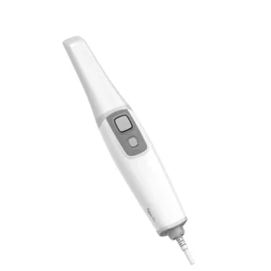
An intraoral scanner captures optical information from a patient’s teeth and oral structures and converts it into a three-dimensional digital model.
This digital information can support digital impressions, laboratory communication, CAD/CAM workflows and other applications within digital dentistry, depending on the scanner, software and clinical workflow.
For dentists, orthodontists, prosthodontists, implantologists, dental clinics and laboratories, an intraoral scanner can replace the physical impression stage in appropriate workflows.
Instead of creating a conventional impression with a tray and material, the clinician captures the required surfaces digitally and reviews the resulting model on a connected computer.
The choice of scanner matters because different systems vary in ergonomics, scanning workflow, output formats, connectivity, software and published performance specifications.
AGKEM’s intraoral scanner category includes the AGKEM PRO V5 Intraoral Scanner, giving dental professionals an option to consider when evaluating digital impression equipment.
An intraoral scanner is a handheld digital device that optically captures the surfaces of teeth and relevant oral structures to create a 3D digital representation.
During scanning, the device is moved over the areas that need to be recorded. The scanner captures a sequence of optical images or data points, and its software processes this information to reconstruct a digital model.
The resulting model is often referred to as a digital impression. Unlike a conventional physical impression, the data exists electronically and can be reviewed, stored and transferred through compatible digital workflows.
A physical impression requires impression material, a tray and subsequent handling. An intraoral scanning workflow instead captures the patient’s dental structures electronically.
This can be particularly useful for clinics and laboratories that already use digital dentistry or are moving towards CAD/CAM-based processes.
An intraoral scanner does not eliminate the need for clinical examination, treatment planning or professional judgement.
It is a data-capture tool that becomes useful when integrated appropriately into the rest of the dental workflow.
An intraoral scanner works by capturing optical data from inside the patient’s mouth and processing that information into a 3D digital model.
The basic process involves:
The scanner tip is placed inside the patient’s mouth and directed towards the teeth and oral structures being recorded.
As the scanner moves, it continuously captures images or optical information from the surfaces being scanned.
The scanning software combines the captured information and identifies the relationship between successive images or data points.
The processed information is assembled into a digital three-dimensional representation.
The dentist can view the developing digital model on the connected computer and identify whether additional areas need to be scanned.
Depending on the scanner and its software, the completed scan can be exported in supported formats for use in compatible laboratory, CAD/CAM or digital dentistry workflows.
The quality of the final digital impression depends on the scanner, software, scanning technique, clinical conditions and the completeness of the captured data.
A scanner should therefore be evaluated as part of the complete workflow rather than judged only by one specification.
An intraoral scanner can support several dental workflows where a digital impression or 3D representation of the dentition is required.
Common applications include:
The scanner does not replace every conventional impression technique or every stage of treatment.
Case requirements, clinical conditions and the laboratory or software workflow should determine whether digital scanning is appropriate.
For laboratories, receiving digital scan data can also reduce the need to handle and transport physical impressions when an open digital workflow is being used.
The benefits of an intraoral digital scanner are primarily related to how dental data is captured, visualised and managed.
Digital scanning does not require the conventional impression material and tray used for a traditional impression.
This can make the impression-capture stage more convenient for many patients, although individual tolerance still varies.
Instead of producing a physical negative impression, the clinician creates a digital representation that can be stored and processed electronically.
The clinician can see the scan developing on screen. Missing or incomplete areas can potentially be identified during the scanning process and recaptured.
Digital scan files can be stored electronically as part of an appropriate patient-record system, reducing the need for physical impression storage.
Digital files can be transferred electronically to compatible laboratory workflows.
This can be useful for clinics working with dental laboratories that already accept digital impressions.
A digital file can be transmitted electronically rather than physically transporting an impression. The actual transfer time depends on the file size, connection and receiving system.
For appropriate cases, digital scanning can reduce dependence on impression trays and conventional impression materials.
Digital data can be duplicated and transferred within permitted clinical workflows without physically reproducing an impression.
An intraoral scanner can act as an entry point into a wider digital workflow involving CAD/CAM, digital design and dental laboratory processes.
These benefits should not be interpreted as a guarantee that digital scanning is clinically superior in every situation.
The appropriate impression method depends on the case and the capabilities of the complete workflow.
Both approaches have a role in dentistry. The main difference is whether the impression information is captured digitally or as a physical impression.
Factor | Intraoral Scanner | Traditional Impression |
Patient experience | No conventional impression material or tray is required | Tray and impression material are used |
Impression process | Optical scanning creates digital data | Physical impression is created |
Digital data | Produces an electronic 3D model | Physical impression must be digitised for a digital workflow |
Storage | Electronic storage | Physical storage |
Laboratory communication | Digital files can be transferred electronically | Physical impression generally needs collection or transport |
Data transfer | Electronic transfer through compatible systems | Physical transfer unless subsequently digitised |
Physical materials | Reduces dependence on conventional impression materials | Requires impression materials and trays |
Workflow | Digital review and export | Physical inspection, handling and transport |
An intraoral scanner can be particularly useful for clinics adopting digital workflows, while traditional impressions remain relevant for situations where digital scanning is unsuitable or unavailable.
Choosing an intraoral scanner for a dental clinic involves more than comparing the headline price. Consider the following factors together.
Review the manufacturer’s published accuracy specifications and understand the conditions under which they are stated.
Accuracy should be considered alongside scanning technique, software and case requirements.
Scanning speed can affect chairside efficiency, but it should not be evaluated separately from ease of use, data quality and workflow.
Because the device is handled during the entire scanning process, weight, grip, dimensions and button placement can influence practical usability.
Consider tip dimensions and shape, particularly when scanning posterior areas or working with patients who have limited oral space.
The scanner tip, handpiece shape and scanning duration can influence the patient experience.
The software should make it practical to capture, review and manage scans. Real-time visualisation can help the operator monitor the developing model.
An open-system scanner can offer greater flexibility when exchanging files with compatible third-party laboratories, design software and CAD/CAM systems.
A closed system may be designed around a specific software ecosystem.
Check which formats the scanner can export and whether they are suitable for your laboratory or design workflow.
Confirm compatibility with the specific CAD/CAM and laboratory systems your clinic already uses or intends to adopt.
Review the connection method and the process for transferring completed scan files to the computer or compatible digital systems.
Ask how software updates are handled and what technical support is available.
Warranty terms, technical support and replacement processes should be reviewed before purchasing any digital equipment.
The purchase price is only one part of the investment. Consider software, accessories, maintenance, support, replacement components and the practical requirements of integrating the scanner into your clinic.
An open-system intraoral scanner allows digital scan data to be exported in supported formats for use across compatible digital workflows, rather than restricting the data to a single proprietary ecosystem.
Common dental scan formats include:
The important point is not simply whether a scanner is described as “open”. Buyers should check the exact formats supported, whether colour information is retained where relevant, and whether the receiving laboratory or CAD/CAM software can use those files.
An open workflow can be particularly useful for clinics that work with multiple laboratories or digital design systems.
Compatibility should always be confirmed with the specific software and laboratory before purchase.
The intraoral scanner price can vary significantly depending on the scanner technology, published performance specifications, software, output formats, workflow model, accessories, warranty, technical support and brand.
When comparing an intraoral scanner price in India, dentists should consider the complete ownership cost rather than comparing only the initial quotation.
Important factors include:
The AGKEM PRO V5 product page currently uses an “Enquire Now” purchase process rather than displaying a fixed public price.
Dental professionals can contact AGKEM for current pricing based on the required configuration.
The AGKEM PRO V5 Intraoral Scanner is the current product featured in AGKEM’s intraoral scanner category.
Its official product page describes a lightweight design, real-time 3D visualisation, multi-angle scanning and an open-system workflow.
The handpiece is published at 160 g without the cable, with dimensions of 243 × 40 × 32 mm.
For clinicians who hold the scanner throughout an appointment, handpiece weight and size are practical considerations when evaluating ergonomics.
AGKEM specifies wired USB 3.0 connectivity, allowing the scanner to be connected to a compatible computer-based setup.
The product page also lists an optical continuous video capture method and 3D video image-mode synchronisation.
The PRO V5 supports multi-angle image capture and includes standard and mini scanner tips. The published standard tip measures 115 × 21 × 16 mm, while the mini tip measures 115 × 17 × 13 mm.
The listed scanning areas are 16 × 14 mm for the standard tip and 12 × 10 mm for the mini tip. These details are relevant when considering posterior access and different intraoral positioning requirements.
For clinics considering an open digital workflow, AGKEM lists STL, PLY with true colour, and OBJ as output formats and states that the scanner is compatible with most design software.
The specific compatibility of a clinic’s existing software or laboratory system should still be confirmed before purchase.
AGKEM publishes scanning accuracy specifications of less than 10 μm for a single crown and less than 20 μm for a full arch.
These are manufacturer-published specifications and should be evaluated in the context of the scanning technique, clinical case and complete digital workflow.
The product page also lists a depth of view of up to 25 mm, 20 FPS image capture, real-time scanning, real-colour technology, quick reset and multi-angle flexible positioning.
Explore the AGKEM PRO V5 Intraoral Scanner
An intraoral scanner can form the first digital data-capture stage of a broader workflow:
Patient → Intraoral Scan → Digital Impression → Digital File → CAD/CAM / Dental Laboratory → Restoration or Appliance
The dentist first captures the required intraoral structures. The software then reconstructs the scan into a digital model, which can be reviewed before the relevant files are exported.
Depending on the clinic and laboratory setup, the digital file may then be transferred to a dental laboratory for design or incorporated into compatible CAD/CAM workflows.
The AGKEM PRO V5 fits into this type of workflow through its published open-system approach and support for STL, PLY and OBJ output.
The key consideration is compatibility. A scanner should be evaluated against the actual workflow used by the clinic and laboratory rather than assuming that every digital scanner works identically with every software platform.
An intraoral scanner may be worth considering when a dental practice or laboratory regularly handles workflows that benefit from digital impressions.
New practices establishing a digital workflow can consider an intraoral scanner alongside other digital equipment.
The decision should be based on procedures, budget, laboratory relationships and expected usage.
Established practices moving from conventional impressions can evaluate whether digital scanning would simplify their existing impression and laboratory workflow.
Digital scans can support orthodontic records and compatible treatment workflows, including suitable clear aligner processes.
Implant-focused practices may use intraoral scanning as part of appropriate restorative and digital treatment workflows.
Digital impressions can support selected crown, bridge and prosthodontic workflows where the equipment and laboratory processes are compatible.
Clinics handling several treatment disciplines may benefit from having a scanner that can serve multiple compatible digital workflows.
Laboratories working with digital impressions can receive electronic scan data from compatible clinics and incorporate it into their digital design processes.
Whether an investment makes sense depends on scan volume, procedures, laboratory workflow, software requirements, operator training and overall cost.
An intraoral scanner is a handheld digital device that captures optical information from a patient's teeth and oral structures to create a 3D digital model.
The resulting digital impression can be reviewed, stored and exported in supported formats for compatible digital dentistry, laboratory and CAD/CAM workflows.
An intraoral scanner is used to create digital impressions and 3D dental models for compatible restorative, orthodontic, prosthodontic, implant-related and laboratory workflows.
It can reduce dependence on conventional physical impressions where digital scanning is appropriate and can make electronic transfer of scan data possible.
An intraoral scanner works by continuously capturing optical data as the handpiece moves over teeth and oral structures.
Software processes multiple captured images or data points and reconstructs them into a three-dimensional model that the operator can view and, where necessary, rescan.
A digital impression is a computer-generated 3D representation of scanned dental structures.
Unlike a conventional impression made with a tray and impression material, it exists as electronic data that can be reviewed, stored and exported in supported formats for compatible digital workflows.
An intraoral scanner is not universally better than a traditional impression because suitability depends on the case and workflow.
Digital scanning can offer electronic data, real-time visualisation, easier transfer and reduced use of impression materials, while conventional impressions remain relevant for situations where digital scanning is not suitable or available.
Intraoral scanner accuracy varies by device, scanning conditions, software, technique and case.
Manufacturers publish accuracy specifications for their systems, but these figures should be assessed in context rather than treated as a guarantee of identical results in every clinical situation.
Operator technique and complete data capture also matter.
There is no single scanning time that applies to every patient or case. Scan duration depends on the scanner, software, operator experience, scan area and case complexity.
AGKEM's PRO V5 page publishes 20 FPS image capture but does not state a single universal scan time for every procedure.
Yes, an intraoral scanner can be used as part of appropriate implant-related digital workflows.
Digital scans can provide the intraoral surface data required for compatible restorative or planning processes.
The scanner is one component of the overall workflow and should be used according to the clinical and technical requirements of the case.
Yes, intraoral scanning can support compatible orthodontic digital workflows.
Three-dimensional digital models can be used for records, treatment planning and selected orthodontic applications.
The specific workflow depends on the scanner, software and orthodontic system being used.
Yes, digital intraoral scans can form part of compatible clear aligner workflows.
The scan provides a three-dimensional representation of the dentition that can be transferred to an appropriate digital planning or laboratory workflow.
Specific software and provider requirements should be confirmed before selecting a scanner.
An open-system intraoral scanner allows scan data to be exported in supported formats for use with compatible external digital systems.
This can provide flexibility when communicating with laboratories or working with different CAD/CAM platforms.
The exact formats and compatibility should always be checked before purchase.
Common intraoral scan formats include STL, PLY and OBJ. The exact formats vary by scanner.
STL generally represents surface geometry, while PLY and OBJ can support additional data depending on how the file is generated.
AGKEM's PRO V5 officially lists STL, PLY with true colour and OBJ output.
STL, PLY and OBJ are different 3D data formats used to transfer digital models. STL is widely used for geometry, while PLY and OBJ can support additional information such as colour depending on the file and software.
The best format depends on the laboratory, CAD/CAM system or application receiving the scan.
Intraoral scanner prices in India vary according to the scanner, software, accuracy specifications, output formats, accessories, ecosystem, support and brand.
AGKEM's PRO V5 product page currently uses an enquiry-based process instead of displaying a fixed public price, so prospective buyers should contact AGKEM for current pricing.
Look at accuracy, scanning workflow, ergonomics, handpiece weight, scanner tips, software, output formats, open or closed workflow, connectivity, data export, support, warranty and total ownership cost.
Most importantly, check whether the scanner fits your actual clinic and laboratory workflow rather than selecting it on specifications alone.
An intraoral scanner can be suitable for a small dental clinic when its digital applications justify the investment.
A clinic should evaluate patient volume, procedures, laboratory relationships, available computer infrastructure, software requirements, operator training and budget before purchasing.
Intraoral scanners can replace traditional impressions in some suitable workflows, but they do not eliminate the need for conventional impressions in every clinical situation.
The decision depends on the case, scanning conditions, clinician preference, laboratory requirements and the capabilities of the scanner and digital workflow.
The AGKEM PRO V5 is a digital intraoral scanner designed for optical 3D scanning and digital impression workflows.
AGKEM publishes a 160 g handpiece, real-time 3D visualisation, multi-angle scanning, wired USB 3.0 connectivity, STL, PLY and OBJ output, and published single-crown and full-arch accuracy specifications.
An intraoral scanner can be part of a broader digital dental setup. Depending on the clinic’s requirements, related AGKEM equipment can include:
When comparing an intraoral scanner, start with the workflow you want to build. Consider the procedures performed, laboratory communication, software requirements, file formats, operator ergonomics, patient experience and long-term ownership cost.
The AGKEM PRO V5 can then be evaluated against those requirements using its published specifications rather than price or marketing claims alone.